
Enhancing MRI Analysis with Prostate.Carcinoma.ai
for PACS and DICOM viewers
Are you ready to enhance prostate MRI analysis with FUSE-AI?
Elevate healthcare excellence with Prostate.Carcinoma.ai, the smart AI-power-up for PACS and diagnostic DICOM viewers. Enhance your viewer with new features and seamlessly integrate the containerized Prostate.Carcinoma.ai analysis module into your trusted diagnostic software.
The time has come to join forces with a robust AI partner - let‘s redefine prostate MRI analysis together
Prostate.Carcinoma.ai enhances healthcare professionals’ efficiency and empowers their workflow:
Freeing radiologist from repetitive tasks and long throughput times
Reducing the burden of time-consuming and mentally exhausting work
Transforming the diagnostic routine into a smooth, smart and stable productive experience

Radiologists’ Benefits
Increased time efficiency
The use of Prostate.Carcinoma.ai helps to mitigate the shortage of skilled medical professionals by automating tedious steps involved in creating manual segmentations, thereby freeing up valuable resources.
More accurate prostate volumetry
More accurate and consistent prostate segmentations in MRI provide more precise prostate volume and PSA density calculations.
Less manual work
Automating manual processes to reduce radiologists' workload can enhance their job satisfaction.
Easy set-up: plug-in solution
The PACS or DICOM Viewer plug-in can be seamlessly integrated into the hospital’s IT system, eliminating the need for additional software installations in everyday clinical practice.
The time has come to join forces with a robust AI partner!
Enhance your viewer with new features and seamlessly integrate the containerized Prostate.Carcinoma.ai analysis module into your trusted diagnostic software.
Elevate healthcare excellence with Prostate.Carcinoma.ai, the smart AI-power-up for PACS and diagnostic DICOM viewers.
provide new AI-based functions to your customer base and address new customers.

benefit from fast integration of new viewer functions without the need for in-house R&D efforts
achieve low risk due to the medical device responsibility of certified software
gain market advantages
profit from a shared revenue model


Book your live demo

Example of combined product features of Prostate.Carcinoma.ai plug-in in Lesion™ software
Fast focus to regions of interests (ROI)
Prostate.Carcinoma.ai automatically segments the prostate, its zones, and conspicuous lesions on all T2w images. A clinical study has shown that this analysis time is reduced by approximately 30%.

Example of combined product features of Prostate.Carcinoma.ai plug-in in LesionTM software
Facilitated volumetry
Achieving more accurate calculations of prostate volume and PSA density may contribute to improving urologists' TRUS-guided biopsies with better treatment options.
Example of combined product features of Prostate.Carcinoma.ai plug-in in LesionTM software
Manual adaption is possible
Radiologists can make manual adjustments directly in the diagnostic software if needed (adapt or delete). Volumes and coordinates are then recalculated automatically.
-
![Prostate MRI without AI]()
Prostate MRI without AI
Figure shows combined product features of Prostate.Carcinoma.ai plug-in in mint Lesion™ software
-
![Automated segmentation of prostate gland]()
Automated segmentation of prostate gland
Figure shows combined product features of Prostate.Carcinoma.ai plug-in in mint Lesion™ software
-
![Automated calculation of PSA density]()
Automated calculation of PSA density
Figure shows combined product features of Prostate.Carcinoma.ai plug-in in mint Lesion™ software
-
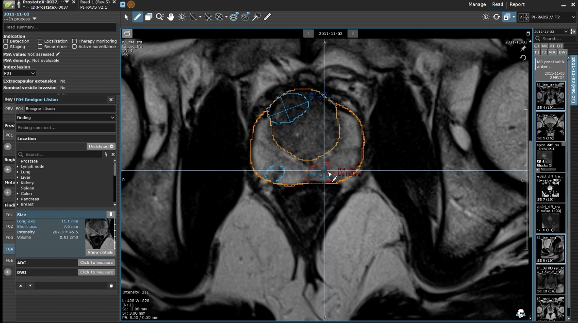
![Automated segmentation of suspicious lesions]()
Automated segmentation of suspicious lesions
Figure shows combined product features of Prostate.Carcinoma.ai plug-in in mint Lesion™ software
-
![Automatic calculation of lesion size and volume]()
Automatic calculation of lesion size and volume
Figure shows combined product features of Prostate.Carcinoma.ai plug-in in mint Lesion™ software


Alexander Cornelius, MD, Head of Urogenital Imaging, Kantonsspital Aarau, Switzerland
“Structured prostate MRI reporting is essential — mint Lesion™ and the FUSE-AI algorithm together significantly boost efficiency and objectivity. I clearly recommend using both systems.“

Felice Burn, MD, Radiologist, Kantonsspital Aarau, Switzerland
“The development of innovative healthcare solutions needs strategic cooperation with competent and motivated partners, who have the capability to face the challenges and the concepts to establish a well digitalized radiology. FUSE-AI understands how to enrich with quality the healthcare market in radiology.“

Mathias Seitel, Head of Product Management, Mint Medical GmbH, Germany
“The shift towards value-based care and evidence-based medicine poses a great challenge for radiology in particular. The deliberate use of new technologies such as Artificial Intelligence is key to making this transition successful. We designed mint Lesion™ just for this purpose: a future-proof radiology workstation that combines contemporary imaging with intelligent reporting tools and thus helps radiologists assume an empowered role in the healthcare system of tomorrow. The collaboration with FUSE-AI and the Cantonal Hospital Aarau complements this strategy. Integrating Prostate.Carcinoma.ai as a plug-in into mint Lesion™ is a logical measure to reach our goal.”


Steffen Rupp, Senior Sales Director LatAm & Europe SW, Mint Medical GmbH, Germany
"Through our partnership with FUSE-AI and the combination of mint Lesion™ with Prostate.Carcinoma.ai, we complement the benefits of structured reporting with those of automatic image recognition. The high standardization, reproducibility, and quality of radiological findings now also coincide with significant efficiency increases in the reporting process," explains Steffen Rupp, Senior Sales Director at Mint Medical. "We are delighted to have found in FUSE-AI an innovative partner to support our users together on the path to data-driven and value-oriented radiology and medicine."
